Meniscus Tear MRI - Getting A Clear Picture Of Your Knee
When your knee starts acting up, feeling a bit off, or maybe even hurting quite a lot, it can really throw a wrench into your daily plans. You might find yourself wondering what's going on inside that important joint. Sometimes, the problem could be something called a meniscus tear, which is a common sort of knee injury. This injury involves the little cushiony bits in your knee, and when they get damaged, it can lead to discomfort, swelling, and just plain trouble moving your leg like you want to, you know?
Figuring out what's causing knee pain is a big step toward feeling better, and that's where modern imaging often comes into play. For something like a meniscus tear, doctors really like to get a good look at what's happening deep inside your knee without having to make any cuts. This is where a special kind of picture-taking, known as a magnetic resonance imaging scan, or MRI for short, becomes incredibly helpful, actually. It gives a very clear view of those internal parts.
This article will talk about why an MRI is such a valuable tool for seeing these tears. We will go over what a meniscus tear is, how an MRI helps spot it, and what kind of things doctors look for on these images. We will also touch on some of the ways medical experts interpret these scans to make sure they get the right diagnosis, so you can get the best care for your knee, more or less.
Table of Contents
- What Exactly is a Meniscus Tear?
- How Does a Meniscus Tear MRI Work?
- Why is Meniscus Tear MRI the Best Choice?
- Who Helps Us Look at Meniscus Tear MRI?
- Christopher Centeno's Ideas on Meniscus Tear MRI
- Dr. Robert LaPrade on Meniscus Tear MRI
- Are There Challenges with Meniscus Tear MRI?
- What to Look For in a Meniscus Tear MRI
What Exactly is a Meniscus Tear?
The menisci are these small, C-shaped pieces of cartilage in your knee, kind of like little shock absorbers between your thigh bone and your shin bone. They're made of a tough, fibrous material and they do a couple of really important jobs. For one thing, they help make your knee stable, keeping everything in place as you move. They also help keep the joint surfaces from rubbing directly against each other, which protects the smooth cartilage that covers the ends of your bones, you know? When these little cushions get damaged, that's what we call a meniscus tear. It's basically a breakdown of that tough, cushiony material.
These tears can happen in a few different ways, and they don't all look the same. Some happen suddenly, perhaps after a twist or a hard landing during sports or even just an awkward step. These are often called acute tears. Other times, they can develop slowly over time, perhaps from repeated stress or just getting older, and those are often called chronic tears, so. The specific way the meniscus gets torn can vary a lot, too, affecting how it feels and what might need to be done about it. It really just depends on how the force hits that knee joint.
Knowing what's going on with these tears, like what kind they are and how bad they might be, is a really big deal for deciding how to help someone get better. This is where getting a clear picture of the inside of the knee becomes super important. It's about seeing the extent of the damage, which then helps doctors figure out the best way to get your knee moving comfortably again and to ease any pain you might be feeling, basically. Without that clear picture, it's a lot harder to make a good plan, you see.
- Sheriff Of Baghdad
- Michelle Obama Transgender
- How Old Is Brigitte Poublon Sherman
- Antonella Ferrari Children
- Megan Skiendiel
How Does a Meniscus Tear MRI Work?
When a doctor wants to get a really good look at a possible meniscus tear, they will often suggest an MRI scan. This particular type of imaging uses a very strong magnetic field, along with radio waves, to create detailed pictures of the inside of your knee. It's a pretty cool process, actually, because it can show both the hard parts, like your bones, and the softer bits, like the ligaments, tendons, and of course, those menisci, very, very clearly. It's considered the best way to spot a torn meniscus because of the amazing detail it provides.
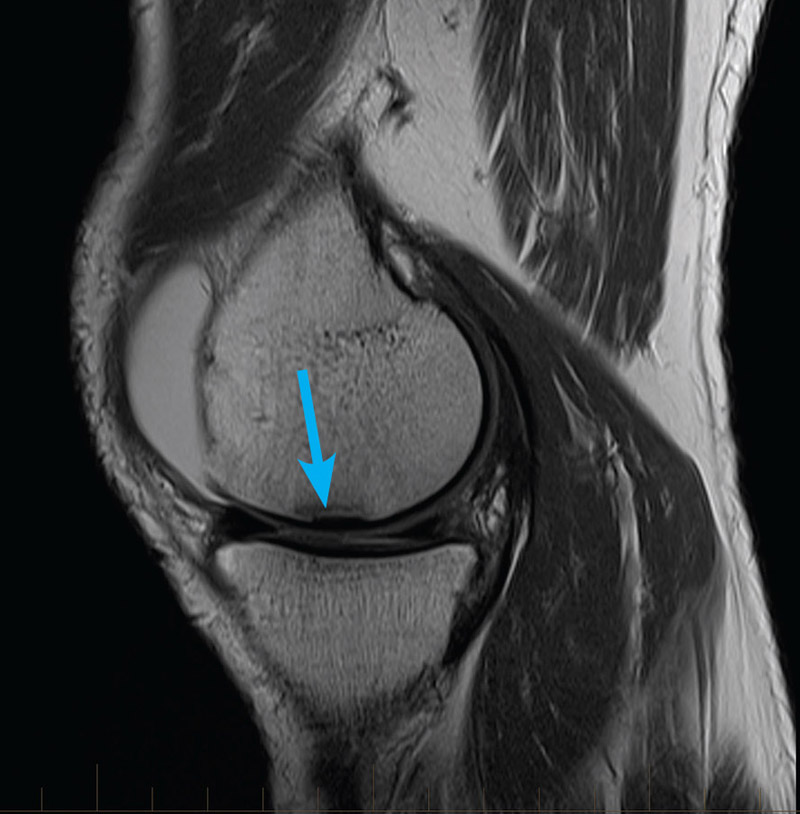
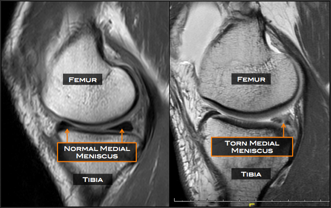
So, how does it help identify a meniscus tear? Well, doctors and radiologists look for certain signs on the MRI images. One of the main things they watch for is a distinct line or a signal that shows up inside the meniscus itself. This signal usually goes all the way from the middle of the meniscus out to one of its edges, either the top or the bottom surface. This kind of line on the scan often means that the meniscus has been sliced or pulled apart in some way, which is what a tear looks like, you know. It's a pretty clear indicator for them.
The way they look at these images has been pretty consistent for a long time, based on a couple of main ideas. They're checking for that unusual signal inside the meniscus, and then they're making sure that signal actually reaches the edge of the meniscus, whether it's the top or the bottom surface. This is particularly true when they use certain types of MRI sequences, which are like different settings on the machine that make specific tissues stand out. This method helps them really pinpoint where the damage might be, so.
Why is Meniscus Tear MRI the Best Choice?
When it comes to figuring out if someone has a meniscus tear, an MRI scan is, by far, the go-to choice for medical professionals. It's incredibly good at what it does, with a very high success rate in picking up these tears. To give you some idea, it can correctly identify a tear up to 93% of the time, and it's also very good at saying when there isn't a tear, being accurate about 88% of the time in that regard, too. These numbers show just how dependable this imaging method is for getting a correct diagnosis, basically.
The reason it's so good is that it provides such clear and detailed pictures. Imagine trying to see a tiny crack in something delicate; you'd want the best possible magnifying glass. An MRI acts like that super magnifying glass for the inside of your knee. It creates these pictures that really show the internal parts of the knee joint, letting doctors see the menisci in great detail. This visual clarity is what makes it so effective at spotting even small tears or other issues that might be causing problems, you know.
Because of this high level of detail, an MRI helps doctors make very accurate diagnoses of meniscus tears. It lets them see the precise location and how big the tear is, which is really important for deciding what kind of help you might need. It's like having a very precise map of your knee, showing exactly where the trouble spots are. This precision is why it continues to be the preferred way to look for these kinds of knee injuries, very, very often.
Who Helps Us Look at Meniscus Tear MRI?
Learning to read an MRI scan for something as specific as a meniscus tear is a skill that takes a lot of practice and knowledge. It's not just about looking at a picture; it's about interpreting what those shadows and lines mean in terms of what's happening inside your body. Luckily, there are experts who have spent a lot of time helping others figure out how to do this. Their insights are really valuable for anyone wanting to get a better handle on what these scans are telling us, so.
These experts often share their wisdom through various means, like videos or detailed explanations. They break down the complex world of medical imaging into more digestible pieces, showing exactly what key features to look for. They might point out common patterns of tears and give practical advice on how to make sense of the MRI results for a knee. It's like having a guide walk you through a complicated map, helping you see the important landmarks that indicate a tear, you know.
Their contributions help ensure that medical professionals, and even curious individuals, can gain a better grasp of what a meniscus tear looks like on an MRI. This shared knowledge is pretty important for making sure that diagnoses are as accurate as possible. It helps everyone involved get a clearer picture of the injury, which then helps guide the next steps for getting the knee healthy again, more or less.
Christopher Centeno's Ideas on Meniscus Tear MRI
One person who has offered some good ideas on how to interpret knee MRIs for meniscus tears is Christopher Centeno. He talks about what it really means to have a meniscus tear, and how that might be different from other knee issues. His work helps people get a better grip on what they're seeing when they look at these scans, and what the findings might mean for someone's knee health. It’s about more than just spotting a line; it’s about what that line represents in terms of the actual injury, basically.
He helps explain the finer points of looking at these images, making it clearer for others to understand the difference between a tear and perhaps just a normal variation in knee anatomy. This kind of detailed instruction is quite helpful for anyone who needs to make sense of an MRI report. It gives them a framework for looking at the images and making good judgments about what's going on with the meniscus, too. His insights are often shared to make the process of reading scans less confusing.
By breaking down the elements of a knee MRI, he helps people see what to focus on when trying to identify a meniscus tear. It's about looking for specific signs that point directly to damage, rather than just guessing. This approach helps reduce the chances of misinterpreting the scan, which is really important for getting the right care. His way of explaining things makes a complex topic feel a bit more approachable, you know.
Dr. Robert LaPrade on Meniscus Tear MRI
Another expert, Dr. Robert LaPrade, a knee surgeon from Minnesota, has also shared valuable information, especially through videos, about meniscus tears and how they appear on MRI. He explains the very specific details of certain tear patterns, even showing examples of what they look like on the actual scans. This kind of visual teaching is very effective because it lets you see firsthand what doctors are looking for, so.
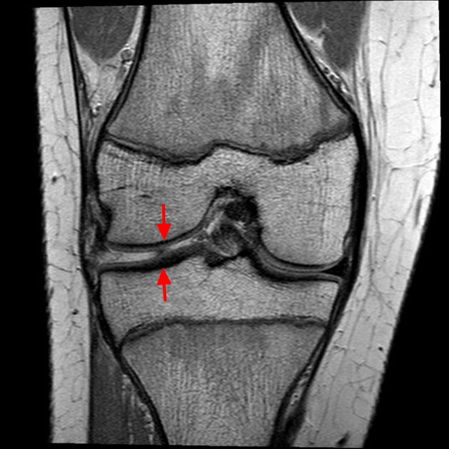
For instance, he discusses how to spot a horizontal cleavage tear, particularly in the medial meniscus, from different angles on an MRI. This type of tear is a specific pattern where the meniscus basically splits horizontally, kind of like a sandwich. Seeing how this looks from various MRI views, like axial, sagittal, or coronal, helps medical professionals get a complete picture of the tear. His explanations are very detailed, showing exactly what to watch out for on each view, you know.
His approach to teaching about these tear patterns is very practical, offering real-world examples that make the subject much clearer. By watching his explanations, people can gain a much better idea of how to identify these specific types of injuries when they're reviewing an MRI. This practical guidance is a big help for improving how accurately tears are diagnosed, which then leads to better care for the patient, honestly.
Are There Challenges with Meniscus Tear MRI?
Even though MRI is really good at finding meniscus tears, there can sometimes be challenges in spotting them. Not every tear jumps out immediately on the scan, and sometimes, they can be a bit tricky to see. This means that the person looking at the MRI needs to be very sharp and know what to look for, even when the tear itself isn't perfectly obvious. It's not always as simple as just seeing a big, clear line, you know.
To avoid making mistakes when trying to figure out if there's a meniscus tear, those who read the MRI scans of the knee need to have a very good grasp of how the menisci are connected inside the knee. They also need to be aware of all the normal ways a meniscus can look that might, at first glance, seem like a tear but aren't. It's like knowing all the little quirks and variations that are perfectly fine, so they don't get confused with actual damage, basically. This deep knowledge helps them make accurate calls.
Sometimes, if a tear is hard to see directly, there are other, indirect signs that can hint at an underlying problem. These are like clues that suggest a tear might be present even if it's not clearly visible. For example, things like a fluid-filled sac near the meniscus, or the meniscus pushing out of its normal spot, or even swelling in the bone marrow nearby, can all make a radiologist suspect that there's a tear hiding. Being aware of these secondary signs is a big part of getting an accurate diagnosis, too.
What to Look For in a Meniscus Tear MRI
When someone is looking at an MRI scan for a meniscus tear, they're typically searching for a specific kind of signal. Usually, a meniscus tear shows up as a line that has a certain brightness, and this line starts from the main part of the meniscus and goes all the way to one of its free edges. It's a pretty distinct sign that the structure has been compromised. This linear signal is one of the most common ways a tear presents itself on these detailed images, so.
There are different ways a meniscus can tear, and each can look a bit different on the MRI. For example, a "radial tear" is one where the meniscus is basically sliced in two, like cutting a pie from the center to the crust. When this happens, the cut edges tend to pull apart from each other. These particular tears can be a bit more serious, especially if the person is younger or if the surrounding cartilage is still in good shape. In those cases, doctors often try to repair the tear, as there's a decent chance of success, you know.
Beyond the common patterns, there's been a lot more learning recently about injuries that happen at the very ends of the meniscus, where it attaches to the bone, or around its outer edge. For decades, most of the focus was on tears in the main body of the meniscus, but now, experts are gaining new insights into these "root" and "peripheral" injuries. This expanded knowledge helps doctors get an even fuller picture of all the possible ways a meniscus can be damaged, which is pretty important for complete care, more or less.
To really get an accurate diagnosis of a meniscus tear from an MRI, it's not just about seeing a line. It also involves knowing the common mistakes that can happen when reading these scans. Being aware of these typical diagnostic errors helps medical professionals avoid them, which then leads to a more precise identification of the tear. It's all about being as careful and informed as possible to make sure the patient gets the right diagnosis, you see.
The menisci, those small, tough structures, are incredibly important for the knee. They don't just help with stability; they also play a big part in keeping the joint's smooth surface healthy over time. So, understanding their structure, how they work, the different ways they can tear, and what happens after surgery is all part of getting a good grasp on knee health. This also means knowing about potential issues that can make diagnosis tricky and recognizing signs that can help improve how accurately tears are spotted. All these pieces come together to help doctors and patients alike deal with knee injuries, basically.
Meniscus Tear Mri Axial View
Medial Meniscus Tear Mri
How To Read Knee Mri Knee Mri Meniscus Tear Mri | Images and Photos finder
